
Medical professionals utilize various forms of body imaging to view the internal structures of their patients. These technological devices reveal tissue inflammation and other internal issues, and doctors use the resulting data created by the images to locate specific diseases and help with the diagnostic process. Because inflammatory conditions cause subtle physical changes within the human body, clear internal pictures are needed by physicians to spot these early abnormalities.
Early Detection
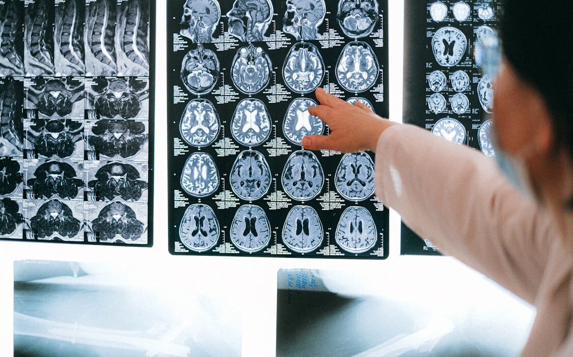
Physicians look for objective signs of disease before severe physical symptoms begin, and modern body imaging scans provide a clear view of early internal structural damage. While standard examinations show external swelling, a magnetic resonance imaging (MRI) scan reveals deeper inflammation before a patient feels pain. Ultrasound machines can also detect early fluid buildup in soft tissues during the initial stages of a medical evaluation. These scans are recorded and viewed later by doctors.
Although blood tests indicate general inflammation, positron emission tomography (PET) scans locate the starting point of the abnormal cellular reaction. Technologists perform these specialized radiological scans to map early biological changes across multiple organ systems. The resulting data helps medical teams create treatment plans, or doctors request additional testing to confirm the initial imaging findings. Extra testing may take the form of blood tests or additional imaging.
Healthcare providers categorize specific inflammatory diseases through the detailed analysis of comprehensive body scans. Since different illnesses attack different organs, a computed tomography (CT) scan helps distinguish between inflammatory bowel disease and appendicitis. Doctors need precise anatomical data to classify the exact condition, and formal imaging reports supply this necessary medical evidence.
Accurate Diagnosis
General physical symptoms mimic many different underlying conditions, but basic imaging can help doctors rule out certain kinds of conditions. By using proper and comprehensive imaging, doctors are able to properly treat and diagnose their patients. Affected anatomical regions are examined by radiologists for specific disease markers associated with localized inflammation. If a patient presents with lung issues, specialized scans show the pattern of respiratory inflammation spreading through the lungs.
Because medical diagnostic teams rely on data, they order various imaging tests to build a diagnostic picture of the patient. An accurate medical diagnosis guides future decisions, including treatment, and the staff updates the patient record with the disease name. These standardized steps formalize the diagnostic phase of routine patient care within the hospital for inflammatory disease diagnosis.
Proper Treatment
Medical clinics base their therapy protocols on the data gathered from these radiological body scans and any additional testing. Doctors monitor tissue changes over an extended period, and they adjust specific medication dosages based on the latest scan results. When a patient undergoes follow-up ultrasound testing, the medical provider determines if the current treatment effectively reduces the internal swelling.
Schedule a Body Imaging Appointment
Body imaging is a useful and accurate method for diagnosing and treating inflammatory diseases. By using these testing methods, doctors can evaluate inflammatory conditions and create individualized treatment plans. Schedule a visit with a radiologist, and discuss receiving a body imaging scan for inflammatory disease.
- The Role of Family Medicine in Managing Family Health Histories
- The Role of CT Scans in Cancer Detection and Monitoring
- The Role of Peptide Therapy in Anti-Aging Treatments
- The Role of Sinus Surgery in Treating Severe Nasal Congestion
- Trusted Home Care Assistance Designed Around Individual Family Needs